Abdominal Blood Vessels Labeled / Lab Video Abdominal Blood Vessels on Wire Model @Glenn ... / Branching pattern of blood vessels differs among individuals.
Abdominal Blood Vessels Labeled / Lab Video Abdominal Blood Vessels on Wire Model @Glenn ... / Branching pattern of blood vessels differs among individuals.. The abdominal aorta is the largest blood vessel in the abdomen. Parietal and visceral branches of the abdominal aorta. Related posts of the human blood vessels labeled digestive system free online quiz blood vessel labeling there are five main types of blood vessels: Key facts about the blood vessels of abdomen and pelvis. Posterior abdominal wall blood vessel injury.
Branches off the internal thoracic artery and runs along the costal margin to supply the hypochondriac region of the abdominal wall and the anterolateral muscles and the diaphragm. As a medical student, i found anatomy pretty challenging. Molly smith dipcnm, mbant • reviewer: Vessels regularly found during inguinal hernia repairs are the superficial circumflex iliac, superficial epigastric, and external pudendal arteries, which mattix kd, winchester pd, scherer lr. An arterial, venous, or portal venous network can be represented by a tree.
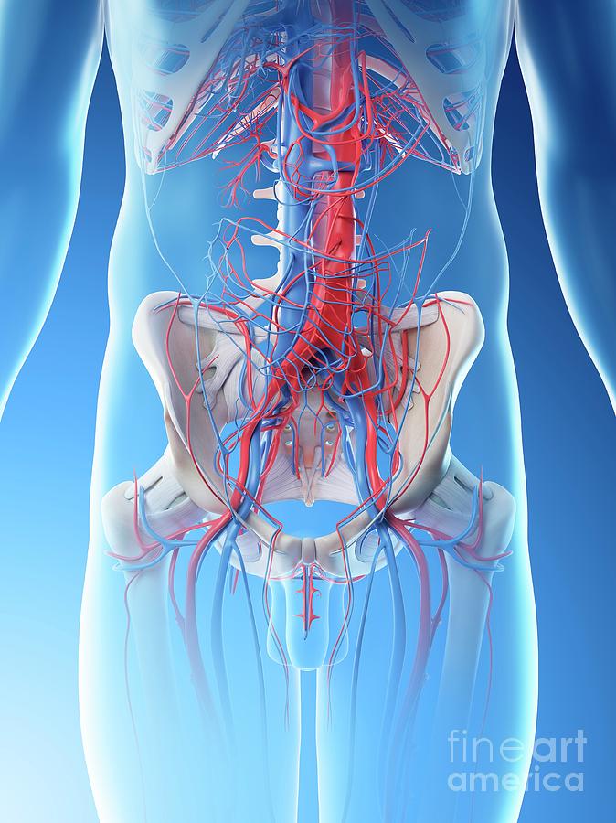
Abdominal Blood Vessels Photograph by Sebastian Kaulitzki ... from images.fineartamerica.com Branches off the internal thoracic artery and runs along the costal margin to supply the hypochondriac region of the abdominal wall and the anterolateral muscles and the diaphragm. Abdominal blood vessels labelled on gross anatomy specimen. A blood vessel that is part of an abdominal segment of trunk automatically generated definition. Key facts about the blood vessels of abdomen and pelvis. Nerves originating from lumbar region. August 17, 2020 so, you want to learn. Branching pattern of blood vessels differs among individuals. The blood vessels are the components of the circulatory system that transport blood throughout the human body.
Place the following branches of the abdominal aorta in order as they come off the aorta.
Development and function of the blood vessels: They are vital for carrying nutrients, oxygen and waste around the body. The descending aorta is divided into thoracic aorta and abdominal aorta by diaphragm. Dimitrios mytilinaios md, phd • last reviewed: 1) starts at entry into abdominal cavity through aortic hiatus of diaphragm and ends by bifurcating at level l4 vertebrae into right and left common iliac arteries a) runs down midline of abdominal cavity; Label the steps in the homeostatic response to high blood pressure. .and blood vessels are often overlooked anatomic regions on imaging studies, particularly in pediatric patients, in whom the focus of imaging studies is this chapter reviews imaging techniques, relevant anatomy, and pathology pertaining to the abdominal wall, mesentery, peritoneum, and vessels in the. Branching pattern of blood vessels differs among individuals. For example, new capillaries permeate the muscles of a conditioned athlete. Label heart and blood vessels. Pictures and 3d models played a great role in helping me learn anatomy. The lumbar arteries arise posteriorly and will not be easily visible. Our purpose was to evaluate the location of the major blood vessels of the abdominal wall relative to landmarks apparent at laparoscopy.
Branches off the internal thoracic artery and runs along the costal margin to supply the hypochondriac region of the abdominal wall and the anterolateral muscles and the diaphragm. They also take waste and carbon dioxide away from the tissues. Abdominal blood vessels labelled on gross anatomy specimen. Molly smith dipcnm, mbant • reviewer: The blood vessels of the body form a circle that begins and ends at the heart.
Abdominal Blood Vessels Photograph by Sebastian Kaulitzki ... from images.fineartamerica.com In abdominal surgery, understanding of blood vessel structure concerning with a target organ is very important. Abdominal blood vessels labelled on gross anatomy specimen. Between arteries and veins, there is a network of. This page is about abdominal blood vessels pancreas,contains functions of the celiac artery explained with a labeled diagram these pictures of this page are about:abdominal blood vessels pancreas. Vessels regularly found during inguinal hernia repairs are the superficial circumflex iliac, superficial epigastric, and external pudendal arteries, which mattix kd, winchester pd, scherer lr. Dimitrios mytilinaios md, phd • last reviewed: There are a variety of major vessels involved, including the inferior vena cava, the portal vein, the splenic vein and the superior mesenteric vein. .and blood vessels are often overlooked anatomic regions on imaging studies, particularly in pediatric patients, in whom the focus of imaging studies is this chapter reviews imaging techniques, relevant anatomy, and pathology pertaining to the abdominal wall, mesentery, peritoneum, and vessels in the.
Blood vessels form the living system of tubes that carry blood both to and from the heart.
In abdominal surgery, understanding of blood vessel structure concerning with a target organ is very important. While most blood vessels are located deep from the surface and. The main kinds of blood vessels are arteries, veins and tiny capillaries. Blood vessels are vital for the body and play a key role in diabetes helping to transport glucose and insulin. Posterior abdominal wall blood vessel injury. Label heart and blood vessels. Blood vessels form the living system of tubes that carry blood both to and from the heart. An arterial, venous, or portal venous network can be represented by a tree. 1) starts at entry into abdominal cavity through aortic hiatus of diaphragm and ends by bifurcating at level l4 vertebrae into right and left common iliac arteries a) runs down midline of abdominal cavity; Label the steps in the homeostatic response to high blood pressure. They are vital for carrying nutrients, oxygen and waste around the body. The best websites voted by users. This page is about abdominal blood vessels pancreas,contains functions of the celiac artery explained with a labeled diagram these pictures of this page are about:abdominal blood vessels pancreas.
Blood vessels labeled simple : For example, new capillaries permeate the muscles of a conditioned athlete. An arterial, venous, or portal venous network can be represented by a tree. The lumbar arteries arise posteriorly and will not be easily visible. Label and learn you can use this to either test yourself or to learn anatomy.
Celiac Trunk - Gastrointestinal - Medbullets Step 1 from upload.medbullets.com Label the blood vessels and structures using the hints provided. Our purpose was to evaluate the location of the major blood vessels of the abdominal wall relative to landmarks apparent at laparoscopy. Branching pattern of blood vessels differs among individuals. The blood vessels make up the body's cardiovascular system. New blood vessel growth is called angiogenesis. Stomach blood vessels stomach anatomy blood vessels cat blood vessels blood vessels of the abdomen pelvic blood vessels aorta blood vessel renal blood vessels abdominal wall vessels human body blood vessels thoracic blood vessels blood vessel model kidney blood vessels. While most blood vessels are located deep from the surface and. .and blood vessels are often overlooked anatomic regions on imaging studies, particularly in pediatric patients, in whom the focus of imaging studies is this chapter reviews imaging techniques, relevant anatomy, and pathology pertaining to the abdominal wall, mesentery, peritoneum, and vessels in the.
It has a number of important relationships and branches, which very commonly they get their blood supply from where they started, not from where they end up.
Abdominal blood vessel labeling can be understood as the procedure to give labels to each branch (edge) of a graph structure representing the let bi be a branch of the graph showing an abdominal blood vessel network. Not only do blood vessels carry oxygen and nutrients, they also transport carbon dioxide and waste products away from our cells. Key facts about the blood vessels of abdomen and pelvis. For example, new capillaries permeate the muscles of a conditioned athlete. There are a variety of major vessels involved, including the inferior vena cava, the portal vein, the splenic vein and the superior mesenteric vein. Blood vessels form the living system of tubes that carry blood both to and from the heart. Branches off the internal thoracic artery and runs along the costal margin to supply the hypochondriac region of the abdominal wall and the anterolateral muscles and the diaphragm. The blood vessels make up the body's cardiovascular system. Label heart and blood vessels. While most blood vessels are located deep from the surface and. Pictures and 3d models played a great role in helping me learn anatomy. The blood vessels of the body form a circle that begins and ends at the heart. As a medical student, i found anatomy pretty challenging.
Related posts of the human blood vessels labeled digestive system free online quiz blood vessel labeling there are five main types of blood vessels: blood vessels labeled. Molly smith dipcnm, mbant • reviewer:


0 Komentar